ADLDpk.org
  • Home
  • About
    • ADLD Team
    • Fast Facts
    • History
    • A Word From Chairman
    • Area Of Operation
    • Aims And Objectives
    • Services offered
    • Mission
    • Plan of operation
    • Executive council
    • Midterm Progress Report
  • Forum
  • Education & Training
    • Practice Guidelines
    • Clinical Methods
    • CME
    • Drugs
  • Blog
  • Gallery
  • Contact
  • More >>
    • Membership
    • Books
    • Endorsements
    • Projects
    • Carrier Opportunities
    • Pedometer
    • Diabetes Risk Calculator
    • Android App

How Scar VT presents - Case presentation by Dr. M. Usman javed

8/19/2017

0 Comments

 
A 74 year old male, resident of Islamabad presented in our ER witrh Multiple episodes of palpitations and dizziness with a blood pressure of 90/50 mmHg during episodes and a heart rate of 160 to 170 bpm. Comorbids include dyslipidemia and Previous Anterior wall MI, 19 years ago Had PCI to LAD at that time.

On presentation his B.P = 125/80mmHg, Pulse = 85 bpm , Afebrile, R.R = 15/min, Spo2 = 99% at room air. Chest was Clear, audible S1, S2 without any added sound or murmur , JVP wasn’t raised, no pedal edema

Baseline Labs:
◦Na = 138 mEq/L
◦K= 4.1 mEq/L
◦Mg = 2.1 mg/dL
◦Ca = 9.4 mg/dL
◦Cr= 0.9 mg/dL
◦CBC – Normal
◦Cardiac Enzymes - Normal
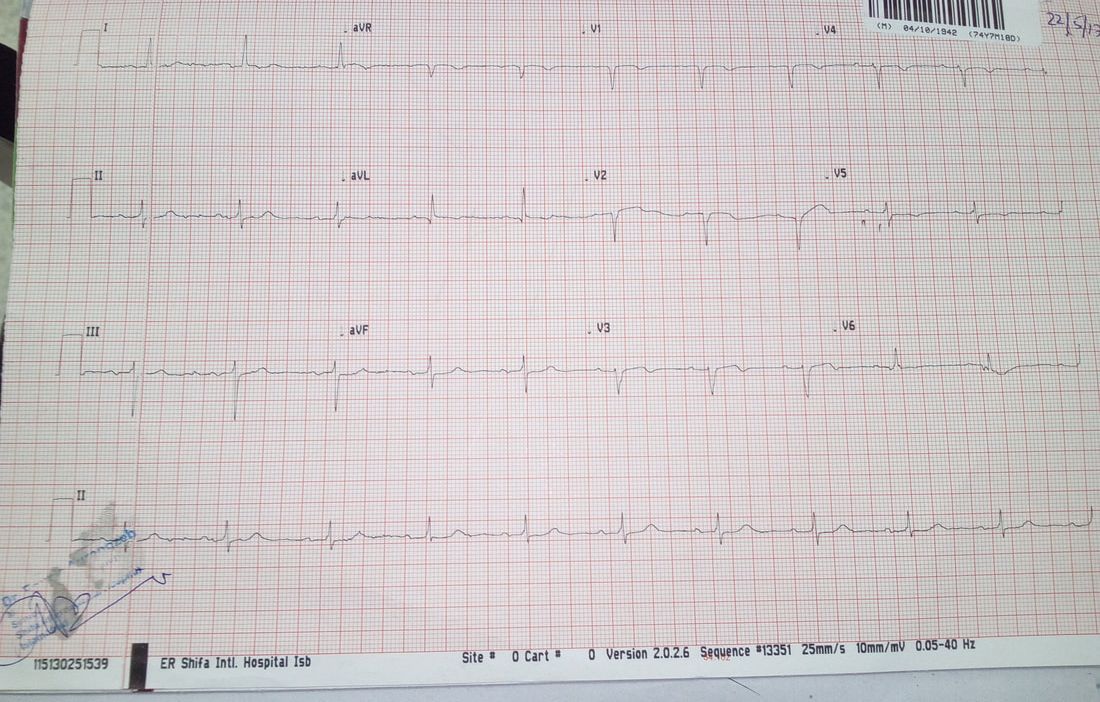
ECG
◦evidence of old anterior wall MI
◦No arrhythmia detected at presentation
Picture
Echocardiography showed a non-dilated left ventricle of 53/35 mm, with moderate systolic dysfunction. LVEF was 30% with hypokinesia of the septum and anterior wall. See the clip below showing parasternal long axis view.
Coronary Angiography showed a patent stent in the proximal left anterior descending artery with mild Instent restenosis. See the clip below showing left coronary system of this patient.
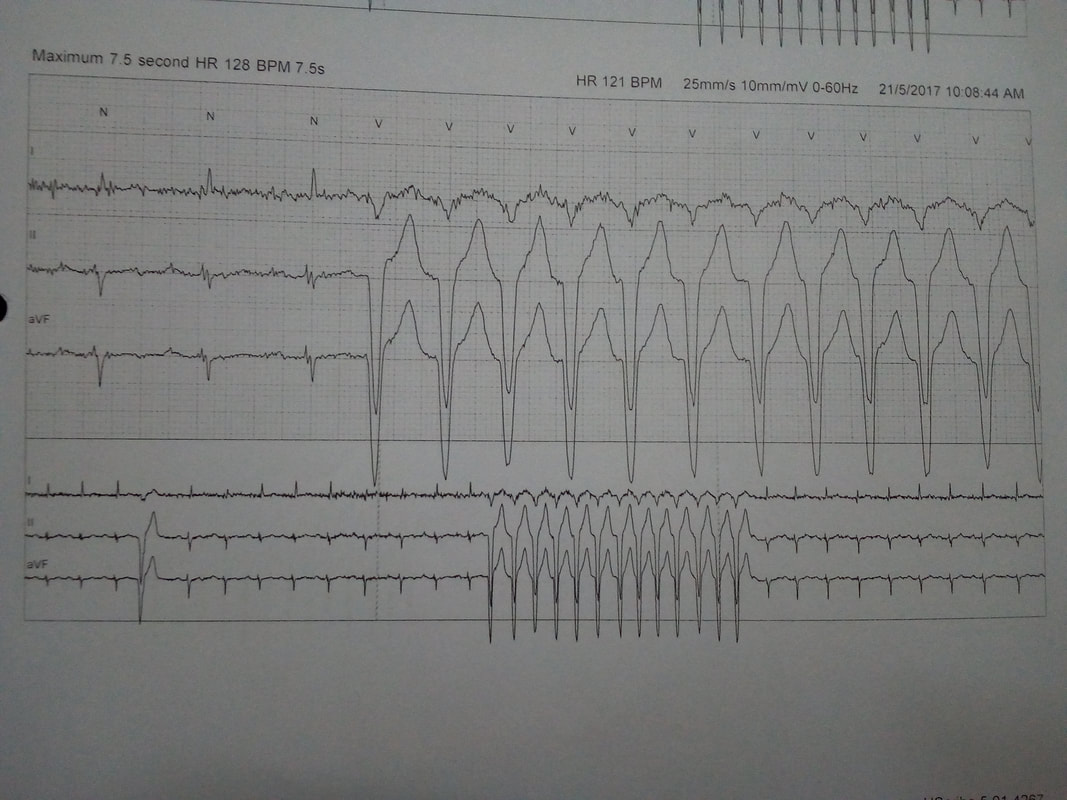
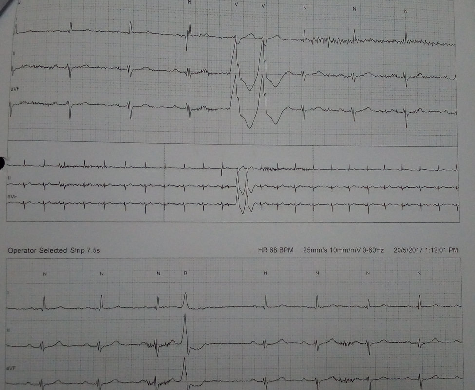
So 48 hour Holter monitoring was advised which showed:
◦Multiple PVCs
◦Runs of sustained VT
Picture
Picture
So it was a typical case of Post MI VT/ Scar related VT. In view of symptomatic VT with depressed LV systolic function, excluding acute ischemia he was managed with maximal medical therapy and implantable cardioverter defibrillator (ICD).
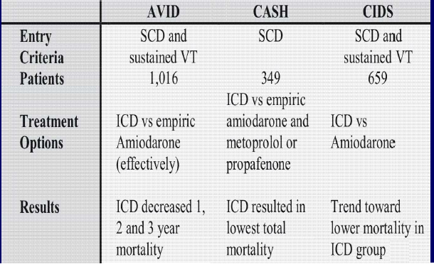
Guidelines:

Picture

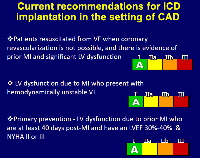
Landmark trials for ICD implantation in post MI VT

Picture
0 Comments

Carcinoid Syndrome ~ CME Presentation by Dr. M Usman Javed

7/25/2016

1 Comment

 
Picture

​Carcinoid Syndrome:

Carcinoid syndrome refers to the array of symptoms that occur secondary to carcinoid tumors. Carcinoid syndrome occurs in approximately 5% of carcinoid tumors. These are diverse group of tumours of neural crest (enterochromaffin cells) origin, by definition capable of producing 5HT (Serotonin). Etiology of carcinoid tumors is not known, but genetic abnormalities are suspected. Reported chromosomal abnormalities include changes in chromosomes, such as numerical imbalances. Manifest when vasoactive substances from the tumors enter the systemic circulation escaping hepatic degradation.

Main  vasoactive substances include:
  • Serotonin
  • Kallikrein (catalyzes the conversion of kininogen to bradykinin which is a powerful vasodilator and bronchoconstrictor)
  • Histamine

If the primary tumor is from the GI tract (hence releasing serotonin into the hepatic portal circulation), carcinoid syndrome generally does not occur until the disease is so advanced that it overwhelms the liver's ability to metabolize the released serotonin.
Picture

Common Sites:

  • Appendix (45%)
  • ileum (30%)
  • Rectum (20%)
  • Other parts of GI tract, ovary, testis, and bronchi (5%)

Signs and symptoms

Signs and symptoms vary greatly and depend on the tumor location and size as well as on the presence of metastases. It may present as
  • Incidental Finding
  • tumor-related symptoms
  • carcinoid syndrome
  • GI tumors can cause appendicitis, intussusception, or obstruction.
  • Hepatic metastases may cause RUQ pain
  • Pellagra
Pellagra is due to niacin deficiency, because tryptophan (which is needed for niacin production) is diverted towards excessive production of serotonin. Leading to Dermatitis, Diarrhea, Dementia.
Serotonin Metabolism

​Diagnosis:

1. Biochemical Markers:

5-hydroxy-indole-acetic acid (5-HIAA)
  • End product of serotonin metabolism
  • Most useful initial test
  • Measured in 24 hour urinary collection
  • Patients with carcinoid syndrome usually excrete >25 mg of 5-HIAA per 24 hour
  • Sensitivity 75% Specificity 100% 

Chromogranin A (CgA) 
  • Present in wall of synaptic vesicles that store serotonin and glucagons
  • Levels correlate with tumour bulk
  • Elevated in 85-100% pts with carcinoid tumour
  • Sensitivity 67.9% Specificity 85.7%

2. Imaging techniques
Conventional Imaging Modalities (CT, MRI, Ultrasound, Endoscopy) are used to locate tumors. Following scans can be performed to diagnose carcinoid tumors.

Octreoscan
  • Initial imaging method  for localization of both primary lesions and metastasis
  • Indium-111 labelled  somatostatin analogues (octreotide ) are used in scintigraphy for detecting tumors expressing somatostatin receptors.
  • Detection rates with octreoscan are about 89%, in contrast to other imaging techniques such as CT scan and MRI with detection rates of about 80%
Meta-iodobenzylguanidine (MIBG) Scintigraphy
  • Used to detect carcinoid and other neuroendocrine tumours
  • Uses 123I- or 131I-labelled MIBG
  • Useful for tumours which do not have somatostatin receptors
Usually on CT scan, a spider-like/crab-like change (Arrow heads) is visible in the mesentery due to the fibrosis from the release of serotonin.
CT scan of a patient with Carcinoid syndrome, a spider-like/crab-like change (Arrow heads) is visible in the mesentery due to the fibrosis from the release of serotonin.

​Treatment:

1. For symptomatic relief of carcinoid syndrome
Octreotide (Sandostatin)
  • somatostatin analogue which decreases the secretion of serotonin by the tumor
  • Initial Treatment with solution: 100-600 mcg/day SC divided q6-12hr; may titrate up to 1500 mcg/day for 2 weeks
  • Maintenance with Suspension (depot injection): 20 mg IM every 4 weeks
2. Surgical resection of tumor
3. Chemotherapy (5-FU and doxorubicin)
4. Endovascular chemoembolization, targeted chemotherapy directly delivered to the liver through special catheters used for patients with liver metastases.
1 Comment

Zika Virus ~ Documentary Video

2/17/2016

0 Comments

 
Zika virus is a member of the virus family Flaviviridae and the genus Flavivirus, transmitted by daytime-active Aedes mosquitoes, such as A. aegypti and A. albopictus. Its name comes from the Zika Forest of Uganda, where the virus was first isolated in 1947. Below is the documentary video about this virus.
0 Comments

Identification Of Basic Structures On Abdominal CT Scan

2/11/2016

0 Comments


Key:

Picture
Picture
Identification of Basic Structures on Abdominal CT Scan: (1) Liver. (2) Spleen (3) Pancreas: head (3+ on images), body, tail (3*), and uncinate process (3-) (4) Gallbladder (5) Right Adrenal Gland (6) Left Adrenal Gland (7) Inferior Vena Cava (8) Aorta (9) Portal Vein (10) Superior Mesenteric Artery (10*) Superior Mesenteric (11) Ascending Colon (12) Descending Colon (13) Transverse Colon (14) Stomach (15) Distal Stomach (16) Right Kidney (17) Left Kidney (18) Inferior Mesenteric Artery (19) Duodenum, 2nd part (19*) Duodenum, 3rd part (20) Left Renal Vein (20*) Left Renal Artery (21) Rt. Renal Vein (21*) Rt. Renal Artery (22) Small Intestine (+) Splenic Artery (x) Common Hepatic Artery (#) Celiac (*) Splenic Vein
 Identification of Basic Structures on Abdominal CT Scan:   (1) Liver.  (2) Spleen   (3) Pancreas: head (3+ on images), body, tail (3*), and uncinate process (3-)  (4) Gallbladder  (5) Right Adrenal Gland  (6) Left Adrenal Gland  (7) Inferior Vena Cava  (8) Aorta  (9) Portal Vein  (10) Superior Mesenteric Artery  (10*) Superior Mesenteric   (11) Ascending Colon     (12) Descending Colon  (13) Transverse Colon  (14) Stomach  (15) Distal Stomach  (16) Right Kidney  (17) Left Kidney  (18) Inferior Mesenteric Artery  (19) Duodenum, 2nd part  (19*) Duodenum, 3rd part  (20) Left Renal Vein  (20*) Left Renal Artery  (21) Rt. Renal Vein (21*) Rt. Renal Artery (22) Small Intestine (+) Splenic Artery (x) Common Hepatic Artery (#) Celiac   (*) Splenic Vein
 Identification of Basic Structures on Abdominal CT Scan:   (1) Liver.  (2) Spleen   (3) Pancreas: head (3+ on images), body, tail (3*), and uncinate process (3-)  (4) Gallbladder  (5) Right Adrenal Gland  (6) Left Adrenal Gland  (7) Inferior Vena Cava  (8) Aorta  (9) Portal Vein  (10) Superior Mesenteric Artery  (10*) Superior Mesenteric   (11) Ascending Colon     (12) Descending Colon  (13) Transverse Colon  (14) Stomach  (15) Distal Stomach  (16) Right Kidney  (17) Left Kidney  (18) Inferior Mesenteric Artery  (19) Duodenum, 2nd part  (19*) Duodenum, 3rd part  (20) Left Renal Vein  (20*) Left Renal Artery  (21) Rt. Renal Vein (21*) Rt. Renal Artery (22) Small Intestine (+) Splenic Artery (x) Common Hepatic Artery (#) Celiac   (*) Splenic Vein
 Identification of Basic Structures on Abdominal CT Scan:   (1) Liver.  (2) Spleen   (3) Pancreas: head (3+ on images), body, tail (3*), and uncinate process (3-)  (4) Gallbladder  (5) Right Adrenal Gland  (6) Left Adrenal Gland  (7) Inferior Vena Cava  (8) Aorta  (9) Portal Vein  (10) Superior Mesenteric Artery  (10*) Superior Mesenteric   (11) Ascending Colon     (12) Descending Colon  (13) Transverse Colon  (14) Stomach  (15) Distal Stomach  (16) Right Kidney  (17) Left Kidney  (18) Inferior Mesenteric Artery  (19) Duodenum, 2nd part  (19*) Duodenum, 3rd part  (20) Left Renal Vein  (20*) Left Renal Artery  (21) Rt. Renal Vein (21*) Rt. Renal Artery (22) Small Intestine (+) Splenic Artery (x) Common Hepatic Artery (#) Celiac   (*) Splenic Vein
 Identification of Basic Structures on Abdominal CT Scan:   (1) Liver.  (2) Spleen   (3) Pancreas: head (3+ on images), body, tail (3*), and uncinate process (3-)  (4) Gallbladder  (5) Right Adrenal Gland  (6) Left Adrenal Gland  (7) Inferior Vena Cava  (8) Aorta  (9) Portal Vein  (10) Superior Mesenteric Artery  (10*) Superior Mesenteric   (11) Ascending Colon     (12) Descending Colon  (13) Transverse Colon  (14) Stomach  (15) Distal Stomach  (16) Right Kidney  (17) Left Kidney  (18) Inferior Mesenteric Artery  (19) Duodenum, 2nd part  (19*) Duodenum, 3rd part  (20) Left Renal Vein  (20*) Left Renal Artery  (21) Rt. Renal Vein (21*) Rt. Renal Artery (22) Small Intestine (+) Splenic Artery (x) Common Hepatic Artery (#) Celiac   (*) Splenic Vein
 Identification of Basic Structures on Abdominal CT Scan:   (1) Liver.  (2) Spleen   (3) Pancreas: head (3+ on images), body, tail (3*), and uncinate process (3-)  (4) Gallbladder  (5) Right Adrenal Gland  (6) Left Adrenal Gland  (7) Inferior Vena Cava  (8) Aorta  (9) Portal Vein  (10) Superior Mesenteric Artery  (10*) Superior Mesenteric   (11) Ascending Colon     (12) Descending Colon  (13) Transverse Colon  (14) Stomach  (15) Distal Stomach  (16) Right Kidney  (17) Left Kidney  (18) Inferior Mesenteric Artery  (19) Duodenum, 2nd part  (19*) Duodenum, 3rd part  (20) Left Renal Vein  (20*) Left Renal Artery  (21) Rt. Renal Vein (21*) Rt. Renal Artery (22) Small Intestine (+) Splenic Artery (x) Common Hepatic Artery (#) Celiac   (*) Splenic Vein
 Identification of Basic Structures on Abdominal CT Scan:   (1) Liver.  (2) Spleen   (3) Pancreas: head (3+ on images), body, tail (3*), and uncinate process (3-)  (4) Gallbladder  (5) Right Adrenal Gland  (6) Left Adrenal Gland  (7) Inferior Vena Cava  (8) Aorta  (9) Portal Vein  (10) Superior Mesenteric Artery  (10*) Superior Mesenteric   (11) Ascending Colon     (12) Descending Colon  (13) Transverse Colon  (14) Stomach  (15) Distal Stomach  (16) Right Kidney  (17) Left Kidney  (18) Inferior Mesenteric Artery  (19) Duodenum, 2nd part  (19*) Duodenum, 3rd part  (20) Left Renal Vein  (20*) Left Renal Artery  (21) Rt. Renal Vein (21*) Rt. Renal Artery (22) Small Intestine (+) Splenic Artery (x) Common Hepatic Artery (#) Celiac   (*) Splenic Vein
 Identification of Basic Structures on Abdominal CT Scan:   (1) Liver.  (2) Spleen   (3) Pancreas: head (3+ on images), body, tail (3*), and uncinate process (3-)  (4) Gallbladder  (5) Right Adrenal Gland  (6) Left Adrenal Gland  (7) Inferior Vena Cava  (8) Aorta  (9) Portal Vein  (10) Superior Mesenteric Artery  (10*) Superior Mesenteric   (11) Ascending Colon     (12) Descending Colon  (13) Transverse Colon  (14) Stomach  (15) Distal Stomach  (16) Right Kidney  (17) Left Kidney  (18) Inferior Mesenteric Artery  (19) Duodenum, 2nd part  (19*) Duodenum, 3rd part  (20) Left Renal Vein  (20*) Left Renal Artery  (21) Rt. Renal Vein (21*) Rt. Renal Artery (22) Small Intestine (+) Splenic Artery (x) Common Hepatic Artery (#) Celiac   (*) Splenic Vein
Picture
0 Comments

How To Check Pulsus Paradoxus - Clinical Signs - Watch Video

11/2/2015

0 Comments

 
  • Definition: On inspiration, a drop in systemic arterial pressure greater than 10mmHg.
  • Measurement:
    1. Inflate the cuff beyond the point where you hear any Korotkoff sounds.
    2. Slowly deflate the cuff until you start hearing any sounds. At the highest BP when you hear sounds intermittently (rather than with every heart beat), note the blood pressure. (This corresponds to the higher systemic blood pressure which is occurring during expiration).
    3. Keep slowly deflating the cuff until you reach the highest BP in which you hear sounds every beat.
    4. The difference between #2 and #3 is the "pulsus paradoxus." If it is >10 mmHg, it is considered significant.
  • DDx: moderate to severe cardiac tamponade, occasionally constrictive pericarditis or COPD exacerbations/asthma attacks.
  • Physiology: Inspiration is performed by decreasing intrathoracic pressure. Because the heart is in the thorax, this pressure is transmitted to the heart and causes a bigger gradient between the pressure in the veins outside of the thorax and the right atrium/ventricle. As such, with each inhalation, more blood flows into the right side of the heart. When this occurs, the interventricular septum subtly bulges into the LV cavity, lowering LV filling, and therefore lowering systemic stroke volume/BP. Because the ventricle can normally also expand outward, this septal shift is usually small, and the difference in the blood pressure is therefore small between inspiration and expiration (<10 mmHg). In states in which the ventricle cannot expand outward (e.g. tamponade) or in which the drop in intrathoracic pressure with inspiration is profound (e.g. status asthmaticus), the septal shift is exaggerated and the difference in BP is larger.
 

Watch Video - Pulsus Paradoxus

0 Comments

Liver transplant in hepatocellular carcinoma - CME - Presentation By Dr. Sidra Zahoor

10/22/2015

0 Comments


Liver transplant in hepatocellular carcinoma

(Topic presentation By Dr Sidra Zahoor - Continuing Medical Education - CME )

Outline
Indications
Milan Criteria
Expanded Milan Criteria (UCSF)
Upto 7 Criteria
MELD Scoring and T staging
Criteria for down staging
LT versus LDT
Pre transplant Evaluation
Procedure
Follow up


Indications: The Milan Criteria
1996, Italy, 48 patients, Mazzafero et al

Single lesion less than 5 cm
Less than 3 lesions none exceeding 3cm
No extrahepatic involvement
No vascular invasion

overall and recurrence-free survivals were 85% and 92%
overall recurrence rate was 8% at 4 years’ follow-up

UCSF-Expanded Milan Criteria
2001

Solitary tumor < 6.5 cm,
or < 3 nodules with the largest lesion < 4.5 cm
and total tumor diameter < 8 cm


Schwartz, Liver Transplantation for Hepatocellular Carcinoma, Gastroenterology.  2004: 127 S268-276.
Vascular invasion

Mc Gill Protocol
Tumors exceeding Milan criteria
Receive 3 TACE treatments at 6 week intervals
Lipiodol,carboplatinum, gelfoam
If patients respond (AFP decreases or tumors shrink, then go on to LT, otherwise continue care)

Upto 7 criteria
HCC with 7 as the sum of the largest tumor (cm) and the number of tumors
Downstaging
LT versus LDT
LDT includes
TACE
RFA
PEI

The published observational studies to date suggest survival following OLT is at least as good as following resection in patients with adequate hepatic reserve.
Liver resection can leave residual liver which is of insufficient size to provide adequate function and as stated before, has the possibility of developing further lesions.
In patients with well-compensated cirrhosis (Child Pugh A) and HCC, the decision whether to resect or transplant remains controversial. However, in the current realm of organ shortage and long waiting times, the decision to resect in this group appears attractive.
If tumor recurrence were to recur than salvage transplantation can be performed.

 An observational series has shown primary OLT to have lower operative mortality, recurrence rates and survival rates
Requirements for listing
UNOS provides a set of specific requirements for listing patients with HCC:
(1)Rough evaluation of the number and size of tumors and to rule out extra-hepatic spread by ultrasound, computed tomography (CT) or magnetic resonance imaging (MRI) plus CT of the chest;
(2) Prelisting biopsy is not mandatory, however patients must have one of the following:
(a) a biopsy confirming HCC;
(b) a vascular blush corresponding to the area of suspected HCC;
(c) an α fetoprotein (AFP) > 200 mg/mL;
(d) an arteriogram confirming a tumor;
(e) a history of local ablative therapy(TACE, RFA, PEI);
(3) Patients with chronic liver disease and a rising AFP > 500 mg/dL can also be listed even in the absence of discrete tumor on imaging studies;
(4) The patient must not be a resection candidate;
(5) Reimaging by CT or MRI every 3 mo is required to ensure continued eligibility for OLT.
Bridging therapy
The “dropout” due to tumor progression whilst waiting for OLT is reported to be at least 20%
This problem has furthered the use of local-regional adjuvant therapy (LDT) whilst awaiting transplantation such as TACE, RFA, and PEI.


The ultimate aim of LDT is to provide complete tumor necrosis in an attempt to halt tumor progression.
Analyses of explant specimens subjected to RFA and TACE have shown complete tumor necrosis rates of 47%-66% and 16%-27%, respectively
A retrospective study looking at tumor necrosis in 61 patients did not find any particular modality of LDT to be superior.
Prognostic Factors
Conforming to the “Milan criteria”, in terms of tumor size and tumor burden, gives rise to 3-4-year recurrence free survival rates of up to 92%

Multivariate analysis has shown these to be the only independent variables predicting patient survival and tumor recurrence. Other biological factors such as tumor grading, microvascular invasion and microsatellites appear to play a role, but within the constraints of the size and number burden.

The histological grade of the tumor can be assessed by lesional biopsy and several authors have recommended this approach. Lesional biopsy does however carry the risk of tumor seeding which has been estimated at approximately 2%

Furthermore, significant histological heterogeneity has been described in large tumors, which limits the utility of needle biopsy
Live donor LT
Living donor liver transplantation (LDLT) has evolved over the past decade, mainly in response to the scarcity of donor livers. Deceased liver donation is particularly scarce in Asia, where organ donation rates are less than 5 donors per million population compared to 10-35 per million in Western countries.

LDLT, in particular right liver transplantation, has dramatically increased the number of potential donors. This can eliminate the problem of long waiting times and ‘dropout’ whilst waiting for an organ because of disease progression.
Furthermore, as there is no direct ‘competition’ from other potential transplant recipients the restrictive criteria on tumor burden can be relaxed somewhat.

Survival rates-same as OLT
Recurrence rates-higher

No consensus guidelines for indications
LDLT carries a risk to the donor during hepatectomy with morbidity and mortality rates of 14%-21% and 0.25%-1%, respectively

Using a scoring system including the measurement of “protein induced by vitamin K absence or antagonist-Ⅱ” (PIVKA-Ⅱ), a Japanese group have achieved a 5-year recurrence rate of only 4.9%. PIVKA-Ⅱ, also known as des-carboxyprothrombin, is an abnormal prothrombin protein found in the serum of patients with HCC and in patients with vitamin K deficiency or on warfarin therapy


Overall survival after olt

Post Transplant Surveillance
Imaging every 3-6 months for 2 years, then annually
AFP if initially elevated-then q3mo for 2 years then q6mo


Download Full Presentation:

liver_transplant_in_hepatocellular_carcinoma.pptx
File Size: 1889 kb
File Type: pptx
Download File

0 Comments

Structure of a rehabilitation program- CME - Presentation By Dr. Sidra Zahoor

10/22/2015

0 Comments


Structure of a rehabilitation program- CME - Presentation By Dr. Sidra Zahoor (Continuing Medical Education)

Joint Diseases
Pre Rehab Assessment
Functional assessment (ie, transfer status, analysis of gait, activities of daily living);
Range of joint motion (ROM) (for all joints);
Muscle strength test (manual or by isokinetic equipment);
Postural assessment; and
Evaluation of respiratory function


Cold/Hot Applications
Cold/hot modalities are the most commonly used physical agents in arthritis treatment. It is well known that cold application is mostly used in acute stages whereas hot is used in chronic stages.

By using heat, analgesia is accomplished, muscle spasm relieved, and elasticity of periarticular structures obtained.
Heat can be used before exercise for maximum benefit. Thermotherapy may be applied as a superficial hot-pack, infrared radiation, paraffin, fluidotherapy, or hydrotherapy. Applications are recommended for 10–20 minutes once or twice a day. Caution is necessary in patients with sensorial deficits and impaired vascular circulation in hands and feet because of burn risk. 

Cold application is preferred in active joints where intra-articular heat increase is undesired. Cold-pack, ice, nitrogen spray, and cryotherapy are different methods of applying cold-therapy.

Levels of destructive enzymes such as collagenase, elastase, hyaluronidase, and protease are affected by the temperature of local joints.
With temperatures of 30° Celsius or lower, effects of these enzymes are negligibly small. Normal intra-articular temperature is 33° Celsius, whereas it may rise up to 36° Celsius in patients with inflammation.
Increasing intra-articular temperature is also related to an increase in collagenase activity and cartilage damage. Despite the inhibition of cell proliferation and metabolic activation within the synovial fluid at 41–42° Celsius, it cannot be used as a therapeutic method because of irreversible joint damage
Electrical Stimulation
Transcutaneous electrical nerve stimulation (TENS) therapy is the most commonly used method.

The highest frequency TENS is the most beneficial, with an analgesia that persists up to 18 hours.

Various studies have reported an increase in hand grip strength after daily application of 15 minutes of TENS and a decrease in pain after using TENS once a week for 3 weeks

Reduction of synovial fluid and inflammatory exudate following TENS application in acute arthritis and suggested that pain relief may be partially explained by this effect. 

Postoperative pain control by TENS therapy following knee joint arthroplasty reduces need for analgesic drugs and hospital stays.
It also has a high placebo effect.
It cannot be used in every painful joint simultaneously, which is a disadvantage in patients with polyarticular involvement. 
Rehabilitative treatment
Joint Protection Strategies
1.Rest and Splinting

Rest and splinting
The joints should be put into rest during the acute stage of the disease.
Bed rest relieves the pain in cases of extensive joint involvement.
It is critical, at this stage, to put the joints into rest at a functional position. Rest position should be as follows: shoulder joint in 45° abduction, both wrist joints in 20° to 30° dorsal flexion, fingers slightly in flexion, hips at 45° abduction without any flexion, knees totally extended, and feet in a neutral position.

Splints may be used to give desired position at rest and functional positioning to the involved active joints. Increased compliance can be gained by offering the patient splints made of soft materials
Orthosis and splinting are used for the following objectives
to diminish pain and inflammation,
to prevent development of deformities,
to prevent joint stress,
to support joints, and
to decrease joint stiffness.

Major factors determining patient compliance to the orthosis are size of the orthosis, the heat generated at the skin by the orthosis, hardness of the parts in contact with the skin, and whether it interferes with functions

Joint stress in the feet may be alleviated by medial arc supporting pad at the sole of the foot and by metatarsal pad. Viscoelastic soles may decrease shock loading occurring at proximal tibia during the gait, by up to 40%.
Compression Gloves
Patients using compression gloves have reported reduced joint swelling and increased well-being.
However, there is no positive evidence regarding improved grip strength or hand functions from using gloves.
Improvement may be provided by using compression gloves for hour intervals or only at night in patients with inflammation in their hands or fingers.
Gentle compression is beneficial because of the containment of joint swelling and subsequent decrease of pain.
Assistive Devices and Adaptive Equipment
Occupational therapy interventions such as assistive devices and adaptive equipment have beneficial effects on joint protection and energy conservation in arthritic patients.
Assistive devices are used in order to reduce functional deficits, to diminish pain, and to keep patients' independence and self-efficiency.
Loading over the hip joint may be reduced by 50% by holding a cane
Massage Therapy
Massage is a commonly used treatment tool that improves flexibility, improves general well being, and can help to diminish swelling of inflamed joints
Pain thresholds both at the massage site and at the knee and ankle decrease after applying oscillatory manual massage to the intervertebral paraspinal region.
Massage is found to be effective on depression, anxiety, mood, and pain.
This finding leads to the question of whether there are some changes in peripheral nociceptive perception and central information in RA. Also, massage decreases stress hormone levels.
Therapeutic Exercise
Muscle weakness in patients may occur because of immobilization or reduction in activities of daily living.
Maintenance of normal muscle strength is important not only for physical function but also for stabilization of the joints and prevention of traumatic injuries.
It may be proposed that exercise therapy has beneficial effects on increasing physical capacity rather than reducing the activity of the disease.

Prior to establishing an exercise program for patients with joint diseases, the following characteristics should be considered:
whether the involvement of the joints is local or systemic,
stage of the disease,
age of the patient,
and compliance of the patient with the therapy.
Duration and severity of the exercise are adjusted according to the patient. ROM exercises, stretching, strengthening, aerobic conditioning exercises, and routine daily activities may be used as components of exercise therapy.

There should be no straining exercises during the acute arthritis.
However, every joint should be moved in the ROM at least once per day in order to prevent contracture.
In the case of acutely inflamed joints, isometric exercises provide adequate muscle tone without exacerbation of clinical disease activity.
Moderate contractures should be held for 6 seconds and repeated 5–10 times each day.

It should be remembered that if isometric exercises are performed in a magnitude of more than 40% of maximum voluntary contraction, they may lead to impairment in blood circulation and fatigue after the exercise.
If the disease activity is low, then isotonic exercises should be performed by using very low weights.
Low-intensity isokinetic knee exercises (by 50% of the maximum voluntary contraction) were reported to be safe and effective in patients

If pain persists more than 2 hours or too much fatigue, loss of strength, or increase in joint swelling occurs after an exercise program, then it should be revised.
Also, walking does not lead to intra-articular pressure increase in healthy subjects but does so in a knee with inflammation and effusion.
Thus, patients with active arthritis should particularly avoid activities such as climbing stairs or weight lifting.
Producing excessive stress over the tendons during the stretching exercises should be avoided.
In sudden stretches, tendons or joint capsules may be damaged.

Finally, in chronic stage with inactive arthritis, conditioning exercises such as swimming, walking, and cycling with adequate resting periods are recommended. They increase muscle endurance and aerobic capacity and improve functions of the patient in general, and they also make the patient feel better.

Patient Education
In patients with joint diseases, sociopsychological factors affecting the disease process such as poor social relations, disturbance of communication with the environment, and unhappiness and depression at work are commonly encountered

Multidisciplinary education with the participation of rheumatologists, orthopedicians, physiotherapists, psychologists, and social workers for patients with arthritis is preferable

In such programs, there is information about benefits and adverse effects of drug therapy, importance of physiotherapy, use of orthosis, psychological coping methods, self-relaxation, and various diets. In addition, patients are taught how to perform the scheduled exercises and how to protect the joints during routine daily life.


Download Full presentation:

structure_of_a_rehabilitation_program.pptx
File Size: 221 kb
File Type: pptx
Download File

0 Comments

Obesity - CME - Presentation By Dr. Sidra Zahoor

10/22/2015

1 Comment


Obesity: (Topic presentation By Dr Sidra Zahoor - Continuing Medical Education - CME )

Step 1:Identifying Patients Who Need to Lose Weight  

Measure height and weight and calculate BMI at annual visits or more frequently.
 
Use the current cutpoints for overweight (BMI >25.0-29.9 kg/m2) and obesity (BMI ≥30 kg/m2) to identify adults who may be at elevated risk of CVD and the current cutpoints for obesity (BMI ≥30) to identify adults who may be at elevated risk of mortality from all causes


Advise overweight and obese adults that the greater the BMI, the greater the risk of CVD, type 2 diabetes, and all-cause mortality.
 
Measure waist circumference at annual visits or more frequently in overweight and obese adults.

Advise adults that the greater the waist circumference, the greater the risk of CVD, type 2 diabetes, and all-cause mortality.  

Need to lose weight
YES – BMI >30 or BMI 25<30 with additional risk factor(s): Weight loss treatment is indicated for 1) obese individuals and 2) overweight individuals with 1 or more indicators of increased CVD risk (e.g., diabetes, prediabetes, hypertension, dyslipidemia, elevated waist circumference) or other obesity related comorbidities  


NO – BMI <25 or BMI 25<30 without additional risk.
Normal weight patients (BMI 18.5<25) should be advised to avoid weight gain.
Patients who are overweight (BMI 25<30), and who do not have indicators of increased CVD risk (e.g., diabetes, prediabetes, hypertension, dyslipidemia, elevated waist circumference) or other obesity-related comorbidities should be advised to avoid additional weight gain  

Step 2:Matching Treatment Benefits With Risk Profiles  

Counsel overweight and obese adults with CV risk factors (high BP, hyperlipidemia and hyperglycemia), that lifestyle changes that produce even modest, sustained weight loss of 3%-5% produce clinically meaningful health benefits, and greater weight losses produces greater benefits.


Sustained weight loss of 3%-5% is likely to result in clinically meaningful reductions in triglycerides, blood glucose, HbA1C, and the risk of developing type 2 diabetes;
Greater amounts of weight loss will reduce BP, improve LDL–C and HDL–C, and reduce the need for medications to control BP, blood glucose and lipids as well as further reduce triglycerides and blood glucose.


At a 3 kg weight loss, a weighted mean reduction in triglycerides of at least 15 mg/dL is observed.
At 5 kg to 8 kg weight loss, low-density lipoprotein cholesterol (LDL–C) reductions of approximately 5 mg/dL and increases in high-density lipoprotein cholesterol (HDL–C) 2 to 3 mg/dL are achieved.
With <3 kg weight loss, more modest and more variable improvements in triglycerides, HDL– C and LDL–C are observed


At a 5% weight loss, a weighted mean reduction in systolic and diastolic BP of approximately 3 and 2 mm Hg respectively, is observed.
• At <5% weight loss, there are more modest and more variable reductions in BP.

Step 3: Determining Recommended goals for weight loss
A realistic and meaningful weight loss goal is an important first step. Although sustained weight loss of as little as 3% to 5% of body weight may lead to clinically meaningful reductions in some CVD risk factors, larger weight losses produce greater benefits. The Panel recommends as an initial goal the loss of 5% to 10% of baseline weight within 6 months.  

Step 4: Determining suitable treatment options
If the weight and lifestyle history indicates that the patient has NEVER participated in a comprehensive lifestyle intervention program, it is recommended that he or she be encouraged to undertake such a program prior to adding adjunctive therapies, as a substantial proportion of patients will lose sufficient weight with comprehensive lifestyle treatment alone to improve health.  


If the patient has been unable to lose weight or sustain weight loss with comprehensive lifestyle intervention and they have a BMI ≥30 or ≥27 with comorbidity, adjunctive therapies may be considered.  

Step 5:Diets for Weight Loss  
Prescribe a diet to achieve reduced calorie intake for obese or overweight individuals who would benefit from weight loss, as part of a comprehensive lifestyle intervention. Any 1 of the following methods can be used to reduce food and calorie intake:  


Prescribe 1,200–1,500 kcal/day for women and 1,500–1,800 kcal/day for men

Prescribe a 500 kcal/day or 750 kcal/day energy deficit

Prescribe one of the evidence-based diets that restricts certain food types (such as high-carbohydrate foods, low-fiber foods, or high-fat foods) in order to create an energy deficit by reduced food intake.


Options

A diet from the European Association for the Study of Diabetes Guidelines, which focuses on targeting food groups, rather than formal prescribed energy restriction while still achieving an energy deficit.
Higher protein (25% of total calories from protein, 30% of total calories from fat, 45% of total calories from carbohydrate) with provision of foods that realized energy deficit.
Higher protein Zone™-type diet (5 meals/day, each with 40% of total calories from carbohydrate, 30% of total calories from protein, 30% of total calories from fat) without formal prescribed energy restriction but realized energy deficit.
Lacto-ovo-vegetarian-style diet with prescribed energy restriction.
Low-calorie diet with prescribed energy restriction.


Low-carbohydrate (initially <20 g/day carbohydrate) diet without formal prescribed energy restriction but realized energy deficit.
Low-fat (10% to 25% of total calories from fat) vegan style diet without formal prescribed energy restriction but realized energy deficit.
Low-fat (20% of total calories from fat) diet without formal prescribed energy restriction but realized energy deficit.
Low-glycemic load diet, either with formal prescribed energy restriction or without formal prescribed energy restriction but with realized energy deficit.
Lower fat (≤30% fat), high dairy (4 servings/day) diets with or without increased fiber and/or low-glycemic index/load foods (low-glycemic load) with prescribed energy restriction.
Macronutrient-targeted diets (15% or 25% of total calories from protein; 20% or 40% of total calories from fat; 35%, 45%, 55%, or 65% of total calories from carbohydrate) with prescribed energy restriction


Mediterranean-style diet with prescribed energy restriction.
Moderate protein (12% of total calories from protein, 58% of total calories from carbohydrate, 30% of total calories from fat) with provision of foods that realized energy deficit.
Provision of high-glycemic load or low-glycemic load meals with prescribed energy restriction.
The AHA-style Step 1 diet (with prescribed energy restriction of 1,500–1,800 kcal/day, <30% of total calories from fat, <10% of total calories from saturated fat).

Step 6: Lifestyle interventions and counselling
Advise overweight and obese individuals who would benefit from weight loss to participate for ≥6 months in a comprehensive lifestyle program that assists participants in adhering to a lower calorie diet and in increasing physical activity through the use of behavioral strategies  


Prescribe on site, high-intensity (i.e., ≥14 sessions in 6 months) comprehensive weight loss interventions provided in individual or group sessions by a trained interventionist  

Electronically delivered weight loss programs (including by telephone) that include personalized feedback from a trained interventionist can be prescribed for weight loss but may result in smaller weight loss than face-to-face interventions.  


Some commercial-based programs that provide a comprehensive lifestyle intervention can be prescribed as an option for weight loss, provided there is peer-reviewed published evidence of their safety and efficacy  


The principal components of an effective high-intensity, on-site comprehensive lifestyle intervention include:
1) prescription of a moderately-reduced calorie diet;
2) a program of increased physical activity; and
3) the use of behavioral strategies to facilitate adherence to diet and activity recommendations  


Comprehensive lifestyle intervention programs typically prescribe increased aerobic physical activity (such as brisk walking) for >150 minutes/week (equal to >30 minutes/day, most days of the week). Higher levels of physical activity, approximately 200 to 300 minutes/week, are recommended to maintain lost weight or minimize weight regain long-term (>1 year).


Use a very low calorie diet (defined as <800 kcal/day) only in limited circumstances and only when provided by trained practitioners in a medical care setting where medical monitoring and high intensity lifestyle intervention can be provided. Medical supervision is required because of the rapid rate of weight loss and potential for health complications.  


Advise overweight and obese individuals who have lost weight to participate long-term (≥1 year) in a comprehensive weight loss maintenance program.  


For weight loss maintenance, prescribe face-to-face or telephone-delivered weight loss maintenance programs that provide regular contact (monthly or more frequent) with a trained interventionist who helps participants engage in high levels of physical activity (i.e., 200-300 minutes/week), monitor body weight regularly (i.e., weekly or more frequent), and consume a reduced-calorie diet (needed to maintain lower body weight).  

Step 7: Pharmacotherapy
Based on expert opinion, the panelists recommend that for individuals with BMI ≥30 or BMI ≥27 with at least 1 obesity-associated comorbid condition who are motivated to lose weight, pharmacotherapy can be considered as an adjunct to comprehensive lifestyle intervention to help achieve targeted weight loss and health goals.  

Step 8: Selecting Patients for Bariatric Surgical Treatment for Obesity  
Advise adults with a BMI ≥40 or BMI ≥35 with obesity-related comorbid conditions who
are motivated to lose weight and
who have not responded to behavioral treatment with or without pharmacotherapy with sufficient weight loss to achieve targeted health outcome goals that bariatric surgery may be an appropriate option to improve health and offer referral to an experienced bariatric surgeon for consultation and evaluation  


For individuals with a BMI <35, there is insufficient evidence to recommend for or against undergoing bariatric surgical procedures.

Advise patients that choice of a specific bariatric surgical procedure may be affected by patient factors, including age, severity of obesity/BMI, obesity-related comorbid conditions, other operative risk factors, risk of short- and long-term complications, behavioral and psychosocial factors, and patient tolerance for risk as well as provider factors (surgeon and facility).  

Options

Laparoscopic adjustable gastric banding (LAGB)
Laparoscopic Roux-en-Y gastric bypass (RYGB)
Open RYGB
Biliopancreatic diversion (BPD) with and without duodenal switch
Sleeve gastrectomy


Step 9: Assess and treat CVD risk factors and comorbidities
Risk assessment for CVD and diabetes in a person with overweight or class I to III obesity includes history, physical examination, clinical and laboratory assessments, including BP, fasting blood glucose, and fasting lipid panel (expert opinion).
A waist circumference measurement is recommended for individuals with BMI 25 <35kg/m2 to provide additional information on risk.
It is not necessary to measure waist circumference in patients with BMI >35 because the waist circumference will likely be elevated and it will add no additional risk information.
The Panel recommends, by expert opinion, using the current cutpoints (>88 cm or >35 in for women and >102 cm or >40 in for men) as indicative of increased cardiometabolic risk. 

Download Full Presentation:

obesity.pptx
File Size: 516 kb
File Type: pptx
Download File

1 Comment

Hepatocellular Carcinoma Management - By Dr. Sidra Zahoor

10/22/2015

1 Comment

 
Hepatocellular carcinoma Management, HCC treatment
HCC Management - By Dr. Sidra Zahoor
CANDIDATE FOR SURGERY
  • Single lesion <5cm (or 6.5cm-UCSF)
  • 3 lesions less than 3cm (or less than 4.5 cm)
  • Ensure absence of mets/Vascular invasions
CANDIDATE FOR DOWNSTAGING
  • Single lesion 5-8cm
  • 2-3 lesions <5cm
  • 4-5 lesions <3cm
  • Total size <8cm
CANDIDATE FOR ABLATION
Single lesion <2cm, not a candidate for surgery

TACE
  • Multifocal, no PVT
  • Beyond milan

For Liver Transplant
Rule Out Contraindications:
  • Mets/PVTT
  • Poor performance status
  • Infection/organ failure
  • Psychosocial issues
1 Comment

    Blog Authors:
    Dr. Saleem Qureshi
    Dr. Musarrat Iqbal
    Dr. Usman Javed

    Archives

    January 2022
    April 2021
    March 2021
    December 2020
    February 2020
    January 2020
    December 2019
    November 2019
    October 2019
    February 2019
    January 2019
    August 2017
    July 2017
    March 2017
    October 2016
    July 2016
    June 2016
    May 2016
    April 2016
    March 2016
    February 2016
    January 2016
    December 2015
    November 2015
    October 2015
    September 2015
    August 2015

    Categories

    All
    ADLD
    Camp Report
    Career
    CDLD
    Clinical Methods
    CME
    Devices
    Diabetes
    Dr Saleem Qureshi
    Dr Suddle
    Dr Usman Javed
    Endorsements
    Imran Abbas
    Insulin Pump
    Medicine
    Motivation
    News
    Photos
    Shoaib Suddle
    Videos
    Zeba Bakhtiar

    RSS Feed

    Picture
    Picture
    Picture
    Picture
    Picture
Powered by Create your own unique website with customizable templates.